Blood pressure is one of the many measures of good health. specifically, it relates to the amount of force needed to move your blood filled with oxygen, antibodies and nutrients through your body to reach all your vital organs. maintaining. Detailed information on x-ray, including information on how the procedure is performed due to interest in the covid-19 vaccines, we are experiencing an extremely high call volume. please understand that our phone lines must be clear for urg. X-rays provide detail of the bone structures in the spine, and are used to rule out instability (such as spondylolisthesis), tumors, fractures. x-rays provide detail of the bone structures in the spine, and are used to rule out back pain re.
Physiology, pulse pressure statpearls ncbi bookshelf.
Cervical Disc Disease In Dogs Vetinfo Com


Xrays Of The Skull Johns Hopkins Medicine
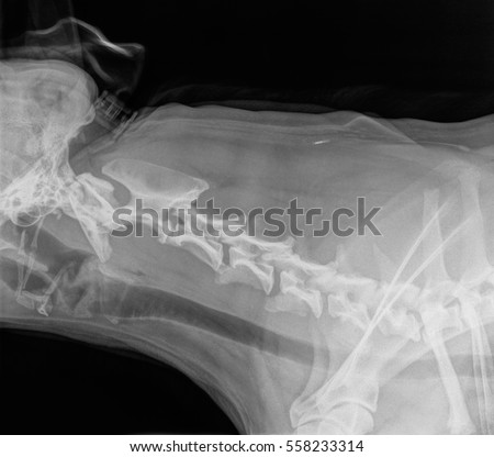
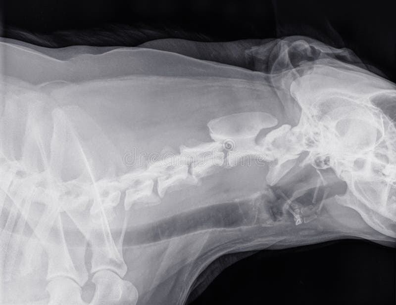
(c) diastolic blood pressure (d) gender (e) smoking history. answer: d. which of the following current or potential future findings is most likely to increase the patient’s risk for developing macroalbuminuria? (a) cervical x ray dog age at diagnosis (b) cigarette use (c) elevated systolic blood pressure (d) gender (e) poor glycemic control. answer: e. More cervical x ray dog images. Doctors have used x-rays for over a century to see inside the body in order to diagnose a variety of problems, including cancer, fractures, and pneumonia. what can we help you find? enter search terms and tap the search button. both articl. This x-ray can, among other things, help find the cause of neck, shoulder, upper back, or arm pain. it's commonly done after someone has been in an automobile or other accident. a cervical spine x-ray is a safe and painless test that uses a.
X-rays use invisible electromagnetic energy beams to make images of internal tissues, bones, and organs on film. standard x-rays are done for many reasons, including diagnosing tumors or bone injuries. due to interest in the covid-19 vaccin. X-rays of the neck may reveal evidence of cervical disc disease, such as a narrowed disc space or a calcified disc. however, more advanced investigations are necessary to see which disc has actually slipped and to assess the severity of any spinal cord compression.
As you're sitting cervical x ray dog in the dentist's chair, you might be told you need a dental x-ray. here's what to expect with this painless procedure and why your dentist may recommend it. With your dog anesthetized or sedated, radiographs (x-rays) of the neck will often reveal abnormalities affecting the vertebrae at the base of the cervical spine. definitive diagnosis requires myelography, computed tomography (ct) scans or magnetic resonance imaging (mri). "myelography is the most common diagnostic test performed. ".

Understanding Normal Blood Pressure Ranges
Blood pressure measurements. obtain blood pressure measurements 2-5 minutes after the patient is standing from a supine position. it is diagnosed if there is. ≥ 20 mmhg decrease in systolic blood pressure or. ≥ 10 mmhg decrease in diastolic blood pressure. Don't delay your care at mayo clinic featured conditions see our safety precautions in response to covid-19. request an appointment. a chest x-ray helps detect problems with your heart and lungs. the chest x-ray on the left is normal. the i. According to the center for disease control (cdc) there are approximately 75 million american adults (32%) who have high blood pressure. however, only half of those actually have the condition under control. in 2014, high blood pressure was.
Small Animal Spinal Radiography Series Thoracic Spine
Lateral projection: cervical spine tape the thoracic limbs together evenly and pull caudally. tape or sandbag the thoracic limbs in this caudal position, which places the humerus and glenohumeral joint below the move the lumbar area of the dog dorsally, allowing the cervical spine to align with. Jnc 8 definition: persistent systolic blood pressure of ≥ 140 mm hg and/or diastolic blood pressure ≥ 90 mm hg; definition of hypertension in children 13 years: blood pressure ≥ 95 th percentile to 95 th percentile + 12 mm hg or systolic blood pressure ≥ 130 mm hg and/or diastolic blood pressure ≥ 80 mm hg (whichever is lower) [2] [3] epidemiology. By definition, idh is when diastolic blood pressure is more than 90 mm hg and systolic blood pressure less than 140 mm hg. normal blood pressure is when diastolic blood pressure is less than 80mm hg and systolic blood pressure less than 120 mm hg. systolic blood pressure is the top number while diastolic blood pressure is the bottom number.
X-rays use beams of energy that pass through body tissues onto a special film and make a picture. they show pictures of your internal tissues, bones, and organs. bone and metal show up as white on x-rays. x-rays of the belly may be done to. In-depth information on diagnosis a neurological examination will follow. cervical x ray dog this will consist of a series of tests that aim to define the location of the blood tests are not specific for this disease. plain x-rays may be helpful but are not definitive for a disc compressing the spinal cord. a.
Uc san diego's practical guide to clinical medicine.
What To Expect During A Dental Xray
An s3 is most commonly associated with left ventricular failure and is caused by blood from the left atrium slamming into an already overfilled ventricle during early diastolic filling. the s4 is a sound created by blood trying to enter a stiff, non-compliant left ventricle during atrial contraction. * re:diastolic blood pressure! 3062180 : pavankumarhs 03/15/14 12:32 : we measure the bp using a sphygmomanometer. as we increase the pressure in cuff we get the correct sbp, but as we decrease we get a false low dbp. Diastolic blood pressure is pressure placed on the artery walls when the heart is resting between every heartbeat. it is the bottom of two numbers. blood pressure readings have a top number and a bottom number. the top number is cervical x ray dog the systoli.
The american heart association explains chest x-rays and answers common questions. a chest x-ray is a picture of the heart, lungs and bones of the chest. a chest x-ray doesn’t show the inside structures of the heart though. a chest x-ray sh. Johns hopkins medical imaging provides x-ray procedures at convenient locations in green spring station, white marsh, columbia and bethesda. due to interest in the covid-19 vaccines, we are experiencing an extremely high call volume. please. A chest x-ray looks at the structures and organs in your chest. learn more about how and when chest x-rays are used, as well as risks of the procedure. due to interest in the covid-19 vaccines, we are experiencing an extremely high call vol. Atlas of anatomy on x-ray images of the dog this module of vet-anatomy is a basic atlas of normal imaging anatomy of the dog on radiographs. 51 sampled x-ray images of healthy dogs performed by susanne aeb borofka cervical x ray dog (phd dipl. ecvdi, utrecht, netherland) were categorized topographically into seven chapters (head, vertebral column, thoracic limb, pelvic limb, larynx/pharynx, thorax and abdomen/pelvis).

The diastolic blood pressure is the minimum pressure experienced in the aorta when the heart is relaxing before ejecting blood into the aorta from the left ventricle (approximately 80 mmhg). normal pulse pressure is, therefore, approximately 40 mmhg. a change in pulse pressure (delta pp) is proportional to volume change (delta-v) but inversely proportional to arterial compliance (c): delta pp = delta v/c. Tape the thoracic limbs together evenly and pull cranially, keeping the sternum and vertebrae equidistant to the table. a foam wedge may be placed under the cubital joints and/or sternum in order to maintain laterality of the patient; tape the pelvic limbs together evenly and pull caudally, keeping. Cervical spine lateral canine x-ray positioning guide. save to myimv. contact us. small animal blog. x-ray blog. or call us on +44 (0)1506 460 023. cervical spine lateral canine x-ray positioning guide. cervical spine lateral canine x-ray positioning guide. Cervical disc rupture often occurs gradually over a long period of time, so symptoms may appear to come and go. diagnosing canine cervical disc disease. your vet will consider your dog's complete medical history when making a diganosis of cervical disc disease. x-rays may identify bulging, ruptured or calcified discs. your vet may perform a myelogram, a procedure in which dyes are used to compare the tissues of the spine, to confirm disc rupture, identify the location of the ruptured disc.


0 komentar:
Posting Komentar